


Magnetic Resonance and Cryogenics
The research of the Nuclear Magnetic Resonance group is oriented primarily towards methodology of localized in-vivo MR spectroscopy, spectroscopic imaging, and quantitative imaging such as diffusometry and relaxometry. These nondestructive, noninvasive and non-ionising measurements are based on the nuclide-selective interaction between magnetic fields produced by the NMR scanner with the atomic nuclei located in the sample volume. The highest sensitivity is achieved with 1H nuclei (protons) and, therefore, the prominent targets are water molecules, which are abundant in biological samples and many materials. In these measurements, the nuclei observed may not only describe the anatomy of the sample interior, but they also act as atomic probes reporting on the nearest environment and processes occurring on the time scale of picoseconds to hours. Besides the ability to identify certain molecules, the acquired data can provide a statistical representation of the fast and random atomic and molecular interactions (manifested by relaxation times, diffusion coefficients, chemical exchange rates and seen as intensity modifications) as well as temporally and/or spatially resolved information on much slower processes (concentration changes, perfusion, flow or motion seen in consecutive images). Quantitative images mapping the spatial distribution of certain well-defined physical properties (relaxation times, diffusion coefficients and directions, flow velocity, temperature etc.) may be obtained and the presence of paramagnetic or superparamagnetic nanoparticles can be detected.
Equipment:
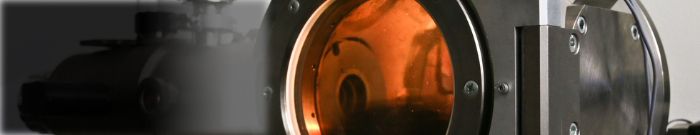
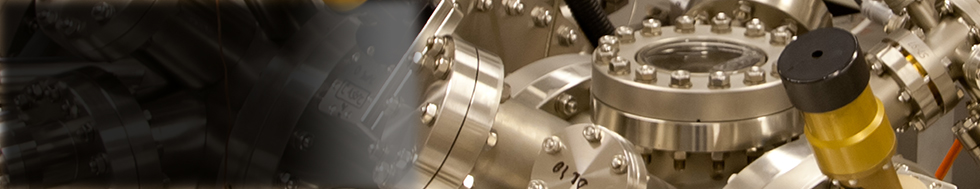
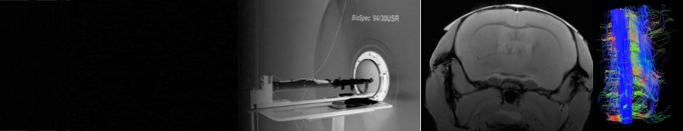
With the MR scanner(s) available in ALISI, virtually all kinds of measurements known from clinical MR scanners can be performed. Thanks to the increased sensitivity resulting from the high magnetic field and small coils, the achievable spatial resolution is much more adequate to the study of small samples. The application sphere is not limited to biomedical imaging; techniques targeting specific problems may be developed.
- MR scanner 4.7 T / 210 mm (proton resonance frequency 200 MHz), optimum sample diameter 40 mm, maximum diameter 100 mm, maximum length about 300 mm; measurable nuclei: 1H, 13C, 19F, 31P, 23Na, 129Xe (immediately), other nuclei negotiable (depending on the availability of RF coils and filters).
- MR scanner 9.4 T / 210 mm (proton resonance frequency 400 MHz, as of mid 2011).
- Physiological functions monitor and isoflurane gas anesthesia unit for small animals (as of 2011).
- RF laboratory.
